Otoacoustic emissions (OAEs) are a valuable tool in audiological assessments, offering insights into cochlear function that complement traditional audiometry. This guide provides clinicians with a comprehensive understanding of OAE measurement, analysis, and interpretation, ensuring accurate and effective patient care.
Learning Objectives
Upon completion of this guide, you will be able to:
- Define criteria for normal otoacoustic emissions (OAEs).
- Identify key measurement factors that can influence OAE test outcomes.
- List the possible outcomes for OAE analysis and their clinical significance.
Agenda
This guide will cover the following topics: the importance of OAEs in clinical practice, recent research on the mechanisms of OAE generation, detailed protocols and guidelines for transient-evoked (TE) and distortion product (DP) OAEs, OAE analysis techniques, factors influencing OAE measurements, and the relationship between OAE findings and audiogram results.
The Cross-Check Principle and OAEs
The cross-check principle, introduced by Jerger and Hayes (1976), emphasizes the importance of using independent measures to validate test results. This principle is particularly relevant in audiology, where behavioral audiometry should be supported by objective measures. OAEs, along with immittance measurements (tympanometry and acoustic reflexes) and auditory brainstem response (ABR), form a crucial part of this test battery. OAEs offer an objective assessment of outer hair cell function, enhancing the accuracy and reliability of audiological evaluations across all age groups.
OAEs are an essential component of a comprehensive test battery, applicable to evaluating hearing from infants to adults. In newborn hearing screenings, combining OAEs with automated ABR yields superior results compared to using either test alone, further demonstrating the cross-check principle in action.
Evidence-Based Practice: OAEs in Pediatric Assessment
OAEs are unequivocally a part of evidence-based audiology for both children and adults. Several clinical guidelines emphasize their importance in pediatric assessment:
- 2007 Joint Committee on Infant Hearing (JCIH) Position Statement
- 2008 American Academy of Audiology Clinical Practice Guidelines: Childhood Hearing Screening
- 2012 American Academy of Audiology: Audiologic Guidelines for the Assessment of Hearing in Infants and Young Children
- 2013 American Academy of Audiology Clinical Practice Guidelines: Pediatric Amplification
- American Academy of Audiology Clinical Practice Guidelines: Otoacoustic Emissions (in progress)
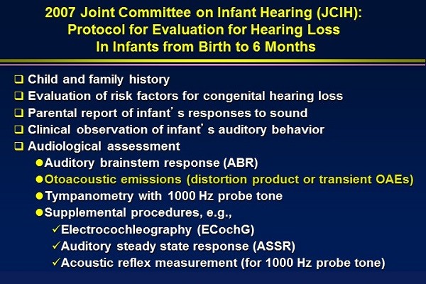
These guidelines highlight the critical role of OAEs in evaluating children, making them a standard of care rather than an optional procedure. The JCIH guidelines (2007) specifically require OAEs for children from birth to six months, reinforcing their necessity in early hearing detection and intervention.
Figure 1. JCIH protocol for evaluation for hearing loss in infants from birth to six months, emphasizing the use of OAEs in early detection.
A Clinician’s Guide to OAE Measurement
Core Principles
This guide operates on the following core principles:
- OAEs are Evidence-Based: OAE measurement is a well-supported and reimbursable procedure for both children and adults.
- OAEs are Underutilized: Many audiologists underutilize OAEs, often employing them as a screening tool rather than a comprehensive diagnostic measure.
- OAEs Require Thorough Analysis: Clinicians should not simply report OAEs as present or absent but should conduct a detailed analysis of the results to extract maximum information.
- OAEs are Valuable for All Ages: OAEs should be routinely recorded in older children and adults, not just young children.
- OAE Replication is Essential: OAE replication is crucial for ensuring accuracy and reliability, minimizing the risk of misinterpreting artifact as genuine OAEs.
- OAEs Provide Unique Information: OAEs offer information beyond what can be obtained from the audiogram, providing a more complete picture of cochlear health.
Understanding OAE Generation
Originally, OAEs were categorized based on recording method (evoked vs. spontaneous) and stimulus type. However, modern research has shifted the focus to the underlying mechanisms of OAE generation.
Reflection Source OAEs
These OAEs, primarily TEOAEs, arise from irregularities along the cochlear partition. Sound travels through the middle ear to the cochlea, and the traveling wave stimulates the outer hair cells. Non-uniform function of these cells, even in normal ears, leads to reflections that are measured as OAEs. The absence of OAEs indicates a lack of outer hair cell function.
Distortion Product OAEs
DPOAEs are generated by the interaction of two frequencies (F1 and F2) that produce a distortion product, typically at 2F1-F2. This distortion is related to the motility of the outer hair cells, where they expand and contract in response to the stimuli. DPOAEs also have a partial reflection component.
Key Differences
TEOAEs and DPOAEs differ significantly in their mechanisms. TEOAEs are reflection-based, while DPOAEs are distortion-based. This is evident in their phase characteristics: TEOAE phase changes with frequency, whereas DPOAE phase remains stable. Employing both TEOAEs and DPOAEs provides a more comprehensive assessment of cochlear function.
Additional Factors in OAE Generation
Beyond hair cells, other cochlear components play crucial roles:
- Prestin: This motor protein is essential for outer hair cell motility.
- Stereocilia: Distortion products are generated by stereocilia, with normal OAEs requiring the tallest stereocilia to be embedded in the tectorial membrane.
- Basilar Membrane Mechanics: Proper basilar membrane function is necessary for OAE generation.
- Stria Vascularis: This highly metabolic area provides blood and oxygen to the cochlea. Issues here can lead to abnormal OAEs despite healthy hair cells.
- Middle and External Ear: While not directly involved in OAE generation, the middle ear’s integrity is crucial for transmitting sound to and from the cochlea. The external ear canal provides the measurement site.
Understanding the roles of Prestin and stereocilia can provide valuable clinical insights, particularly when encountering patients with specific cochlear issues.
Fundamental Steps for OAE Measurement
Before recording OAEs, four fundamental steps must be followed:
- Otoscopy: Always perform otoscopy to ensure the ear canal is clear of obstructions like cerumen or vernix in newborns.
- Middle Ear Measurements: Assess middle ear function using tympanometry and acoustic reflexes.
- Stimulus Calibration: Calibrate the probe at the beginning of each clinical day and verify intensity levels for each patient.
- Monitor Noise Levels: Ensure noise levels in the ear canal are below the 95th percentile for a normal subject.
Middle Ear Considerations
Middle ear measurements are especially critical in infants, as per the JCIH guidelines. Absent acoustic reflexes and abnormal tympanograms indicate potential middle ear issues.
For patients with ventilation tubes, OAE results can vary. Absent OAEs may suggest a non-functional tube, residual middle ear problems, or underlying cochlear issues. Conversely, normal OAEs can be present with patent and functional tubes.
Figure 2. DPOAEs in a child with patent tympanostomy tubes, demonstrating that normal OAEs can be present with functional tubes.
Stimulus Calibration Best Practices
Calibration ensures accurate stimulus presentation. The potential for calibration errors, especially at high frequencies, necessitates careful monitoring. Constructive or destructive interference in the ear canal can affect calibration, so consistent intensity levels are essential throughout the test.
Noise Level Management
Minimizing noise is crucial for reliable OAE measurements. Reduce ambient noise by closing doors, turning off equipment, and ensuring a tight probe fit. In severe cases, consider avoiding low-frequency OAE measurements, as noise levels tend to be highest in that range. Physiological noise can be minimized by instructing older children and adults to remain still and quiet.
Figure 3. Comparison of DPOAE recordings with a poor noise floor (top) and an acceptable noise floor (bottom).
OAE Measurement Tips
TEOAEs offer less noise control but higher frequency resolution, while DPOAEs allow precise frequency selection for noise management. In diagnostic settings, using both TEOAEs and DPOAEs is beneficial.
For diagnostic DPOAEs, record at least five to eight frequencies per octave to capture subtle cochlear abnormalities. This provides detailed frequency information, crucial for identifying issues like noise-induced hearing loss or tinnitus.
Figure 4. Including five to eight frequencies per octave allows for the detection of subtle dips in OAE responses due to cochlear abnormalities.
Remain actively involved during OAE measurements, addressing any issues promptly. Critical review of results during the procedure is essential.
Figure 5. Simple troubleshooting steps for OAE measurements, including optimizing probe fit, reducing noise, and verifying stimulus intensity.
Creating Effective Test Protocols
Customize test protocols to suit the majority of patients. Here are some DPOAE protocol recommendations:
General Diagnostic DPOAE
- Intensity: 65/55 dB
- F2:F1 Ratio: 1.20
- F2 Frequencies: 500 Hz to 8000 Hz (five to eight frequencies per octave)
This protocol suits any cooperative patient.
Specialized Protocols
- Ototoxicity Monitoring/Noise-Induced Hearing Loss: Focus on frequencies from 2000 to 10,000 Hz.
- Meniere’s Disease/Low-Frequency Loss: Use low-frequency OAEs to assess outer hair cell function in these conditions.
Screening Protocol
- Limit frequencies to those critical for speech understanding.
- Use fewer frequencies per octave.
- Replicate results for verification.
Figure 6. DPOAE test protocols for clinical applications, including general diagnostic, ototoxicity monitoring, and screening protocols.
OAE Analysis Criteria
- Noise Floor: Ensure noise floors are adequately low before analysis.
- Frequency Range: Analyze all frequencies to obtain a comprehensive view of cochlear function.
- Repeatability: Verify that OAEs are repeatable to ensure reliability.
Analysis Steps
- OAE-to-Noise Floor Difference: Is there at least a 6 dB difference between the OAE amplitude and the noise floor?
- Normative Comparison: Compare OAE findings to normative data for the specific equipment.
TEOAE Analysis
- Stimulus Adequacy: The stimulus should have a distinct temporal waveform and a broad spectrum.
- Noise Levels: Ensure noise is adequately low.
- Stimulus Stability: High stimulus stability (e.g., 98%) indicates consistent stimulus presentation.
- Response Waveform: Examine the response waveform, noting the timing of responses from different cochlear regions.
- Correlation: Assess correlation between alternating stimulus responses.
- Spectrum: Evaluate the spectrum of the response above the noise floor.
- Signal-to-Noise Ratio (SNR): Measure the amplitude of the OAE compared to the noise.
DPOAE Analysis
- Stimulus Quality: Ensure strong, well-calibrated stimuli.
- Replication: Assess repeatability of OAEs.
- Normative Data: Compare OAE amplitudes to normative data, typically represented by percentile lines.
- Noise Floor: Evaluate the noise floor to ensure accurate measurements.
Figure 7. Analysis of DPOAEs, showing the relationship between F1, F2, and the DPOAE frequency, as well as the calibration and intensity properties.
Figure 8. DPOAEs with poor high-frequency replication, low-frequency absence, and a dip at 1000 Hz, providing valuable diagnostic information.
Figure 9. Abnormal screening DPOAEs, emphasizing the importance of thorough analysis beyond a simple pass/fail determination.
Possible OAE Outcomes
The three possible OAE outcomes are:
- Normal: OAEs fall within the normal range across all frequencies.
- Present but Abnormal: OAEs are present but fall outside the normal range at certain frequencies.
- Absent: No OAEs are present across all frequencies.
Report OAE findings by indicating which of these outcomes is observed across the frequency region from 500 Hz to 8000 Hz.
Factors Influencing OAE Interpretation
Several factors do not influence OAEs, including the time of day, body position, anesthetic agents (with normal middle ear status), and the patient’s attention or cognitive status.
Figure 10. Non-factors in OAE interpretation, highlighting the objectivity of OAE measurements.
However, factors that do influence OAEs include ear canal status, noise levels, middle ear status, and age (in young children but not in older adults with normal hearing).
Figure 11. OAE from an infant, demonstrating the influence of age and ear canal size on OAE amplitudes.
When OAEs are absent, identify the cause: middle ear issues, external ear canal obstructions, or cochlear dysfunction.
OAEs and Audiometric Findings
OAEs validate audiograms and detect cochlear problems that audiograms may miss. Even with up to 20% hair cell damage, audiograms may remain normal, whereas OAEs will be abnormal. OAEs are more sensitive than audiograms in detecting early cochlear dysfunction.
Changes in OAEs can indicate potential hearing problems before they are evident on the audiogram, allowing for early intervention.
Outer Hair Cell Abnormalities and Loudness Recruitment
OAEs are essential for normal hearing of soft sounds. Without OAEs, loudness recruitment occurs, where there is an abnormal growth of loudness.
Abnormal OAEs are associated with abnormal tuning curves, suggesting processing issues even when the audiogram is normal.
OAEs are a pre-neural response, produced by outer hair cells before the synapse with the auditory nerve. Therefore, normal OAEs can occur in patients with hearing loss, indicating potential auditory neuropathy.
OAE and Audiogram Discrepancies
- Agreement: Normal OAEs and normal audiogram usually indicate normal cochlear function.
- OAEs Abnormal, Audiogram Normal: This suggests subtle outer hair cell dysfunction or minor middle ear problems not affecting the audiogram.
- OAEs Normal, Audiogram Abnormal: This could be due to technical issues, inner hair cell dysfunction, or neural problems such as neuropathy.
OAEs are invaluable in detecting false hearing loss, particularly in children. Normal OAEs in a patient presenting with hearing loss on the audiogram raise suspicion of malingering.
Conclusion
OAEs are a vital part of the audiological test battery, enhancing our ability to detect auditory problems at the earliest stages. Early detection leads to more accurate diagnoses and more effective interventions, improving patient outcomes.


