A comprehensive is essential for anyone delving into anatomy and physiology, offering a structured approach to understanding the skeletal system. At CONDUCT.EDU.VN, we provide detailed resources to simplify complex anatomical concepts and enhance your learning experience, making the study of bone anatomy and physiology accessible to students, professionals, and educators alike. This in-depth exploration will cover every aspect of the skeletal system, from its functions and classifications to the intricacies of bone structure and joint mechanics, ensuring you have a robust understanding of skeletal anatomy and physiology.
1. Decoding the Skeletal System: Essential Functions
The skeletal system isn’t just about providing structure; it’s a dynamic framework supporting and enabling numerous bodily functions. Here’s a detailed look at its critical roles:
- Support: Bones act as the body’s internal scaffolding, supporting soft tissues and organs. The vertebral column, for instance, supports the trunk, while the rib cage protects the thoracic cavity.
- Protection: Bones shield vital organs from injury. The skull safeguards the brain, the rib cage protects the heart and lungs, and the vertebrae protect the spinal cord.
- Movement: Bones serve as levers for skeletal muscles, facilitating movement. Muscles attach to bones via tendons, enabling a wide range of motions.
- Storage: Bones store essential minerals like calcium and phosphorus, crucial for various physiological processes. Bone marrow also stores fats.
- Blood Cell Formation: Hematopoiesis, the production of blood cells, occurs in the red bone marrow found in certain bones.
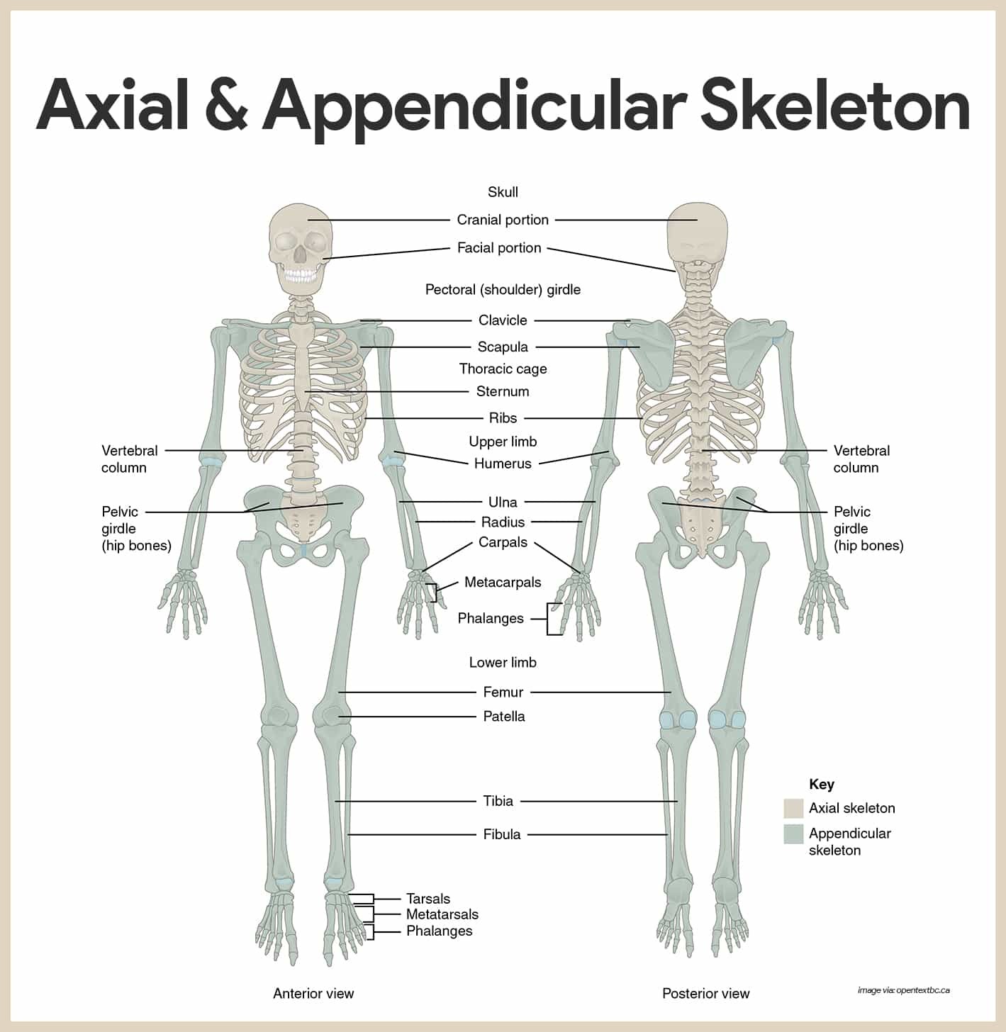
2. Axial vs. Appendicular Skeleton: A Detailed Comparison
The skeletal system is divided into two main parts: the axial and appendicular skeletons. Each has unique components and functions.
- Axial Skeleton: This includes the bones of the skull, vertebral column, and thoracic cage. It forms the central axis of the body, protecting internal organs and providing support.
- Appendicular Skeleton: This consists of the bones of the limbs (upper and lower) and the girdles (shoulder and pelvic) that attach the limbs to the axial skeleton, enabling movement and interaction with the environment.
3. Classifying Bones: Shape and Structure
Bones are classified into five main types based on their shape and structure:
- Long Bones: Longer than they are wide, with a shaft and two ends. Examples include the femur, tibia, fibula, humerus, radius, and ulna. They are primarily found in the limbs and function as levers for movement.
- Short Bones: Cube-shaped, with a spongy bone core covered by a thin layer of compact bone. Examples include the carpals and tarsals, providing stability and support with limited motion.
- Flat Bones: Thin, flattened, and often curved. They consist of two layers of compact bone sandwiching a layer of spongy bone. Examples include the skull bones, ribs, and sternum, providing protection and broad surfaces for muscle attachment.
- Irregular Bones: Complex shapes that don’t fit into the other categories. Examples include the vertebrae and some skull bones like the sphenoid and ethmoid, providing specialized functions.
- Sesamoid Bones: Embedded in tendons, protecting them from stress and wear. The patella (kneecap) is the most prominent example.
4. Long Bone Anatomy: A Comprehensive Overview
Understanding the structure of a long bone is crucial for grasping skeletal physiology. Here’s a detailed breakdown:
4.1. Gross Anatomy
- Diaphysis: The long, cylindrical shaft of the bone, composed mainly of compact bone, providing strength and support.
- Epiphyses: The expanded ends of the bone, composed of spongy bone covered by a thin layer of compact bone. They articulate with other bones at joints.
- Periosteum: A tough, fibrous membrane covering the outer surface of the diaphysis. It contains blood vessels, nerves, and cells responsible for bone growth and repair. Sharpey’s fibers secure the periosteum to the underlying bone.
- Articular Cartilage: A smooth, hyaline cartilage covering the articular surfaces of the epiphyses, reducing friction and absorbing shock at joints.
- Epiphyseal Line/Plate: The epiphyseal plate (growth plate) is a hyaline cartilage plate in the metaphysis at each end of a long bone. It is the part of a long bone where bone growth occurs. The epiphyseal line is the remnant of the epiphyseal plate after bone growth has ceased.
- Medullary Cavity: A hollow space within the diaphysis, filled with yellow bone marrow (adipose tissue) in adults and red bone marrow (hematopoietic tissue) in children.
- Bone Markings: Surface features of bones that serve as sites for muscle attachment, passage of blood vessels and nerves, or formation of joints. These include projections (processes) and depressions (cavities).
4.2. Microscopic Anatomy
- Osteocytes: Mature bone cells located within lacunae (small cavities) in the bone matrix.
- Lacunae: Small cavities in the bone matrix that house osteocytes.
- Lamellae: Concentric layers of bone matrix surrounding a central canal.
- Central (Haversian) Canals: Channels running longitudinally through the bone, containing blood vessels and nerves.
- Osteon (Haversian System): The basic structural unit of compact bone, consisting of a central canal and surrounding lamellae.
- Canaliculi: Tiny channels radiating from the lacunae, connecting osteocytes to each other and to the central canal, allowing for nutrient and waste exchange.
- Perforating (Volkmann’s) Canals: Channels running perpendicular to the central canals, connecting them to each other and to the periosteum, providing pathways for blood vessels and nerves.
5. The Axial Skeleton: Detailed Exploration
The axial skeleton forms the core of the body, providing protection and support.
5.1. Skull: Cranium and Facial Bones
The skull is composed of the cranium and facial bones.
- Cranium: Encloses and protects the brain. It consists of eight bones:
- Frontal Bone: Forms the forehead and the upper part of the eye sockets.
- Parietal Bones: Form the sides and roof of the cranium.
- Temporal Bones: Form the lower sides of the cranium and contain the ear structures. Important markings include:
- External Acoustic Meatus: The ear canal.
- Mastoid Process: A bony prominence behind the ear, serving as an attachment site for muscles.
- Styloid Process: A slender projection below the ear, serving as an attachment site for ligaments and muscles.
- Zygomatic Process: Articulates with the zygomatic bone to form the zygomatic arch (cheekbone).
- Occipital Bone: Forms the back of the cranium and contains the foramen magnum.
- Foramen Magnum: A large opening through which the spinal cord passes.
- Occipital Condyles: Articulate with the atlas (C1 vertebra).
- Sphenoid Bone: A butterfly-shaped bone that forms part of the base of the cranium, the eye sockets, and the nasal cavity. Key features include:
- Sella Turcica: A saddle-shaped depression that houses the pituitary gland.
- Optic Canal: Allows passage of the optic nerve.
- Ethmoid Bone: Located between the eye sockets, forming part of the nasal cavity and the eye sockets. Notable structures include:
- Crista Galli: A vertical projection to which the dura mater (brain covering) attaches.
- Cribriform Plate: A perforated plate that allows passage of olfactory nerves.
- Superior and Middle Nasal Conchae: Scroll-like projections that increase the surface area of the nasal cavity.
- Facial Bones: Form the face and provide attachment points for facial muscles. They consist of fourteen bones:
- Maxillae: Form the upper jaw and contain the upper teeth.
- Palatine Bones: Form the posterior part of the hard palate.
- Zygomatic Bones: Form the cheekbones.
- Lacrimal Bones: Small bones in the medial eye sockets, containing the lacrimal duct.
- Nasal Bones: Form the bridge of the nose.
- Vomer: Forms the inferior part of the nasal septum.
- Inferior Nasal Conchae: Scroll-like bones in the nasal cavity.
- Mandible: Forms the lower jaw and contains the lower teeth.
5.2. Hyoid Bone
The hyoid bone, though not part of the skull, is closely associated.
- Location and Function: Located in the neck, it supports the tongue and provides attachment points for muscles involved in swallowing and speech.
- Unique Feature: It does not articulate directly with any other bone.
5.3. Vertebral Column (Spine)
The vertebral column supports the body and protects the spinal cord.
- Structure: Consists of 26 vertebrae separated by intervertebral discs.
- Regions:
- Cervical Vertebrae (7): Located in the neck.
- Atlas (C1): Supports the skull, allowing for nodding movements.
- Axis (C2): Allows for rotational movements of the head.
- Thoracic Vertebrae (12): Located in the upper back, articulating with the ribs.
- Lumbar Vertebrae (5): Located in the lower back, supporting the weight of the upper body.
- Sacrum: A triangular bone formed by the fusion of five vertebrae, articulating with the hip bones.
- Coccyx: The tailbone, formed by the fusion of three to five vertebrae.
- Cervical Vertebrae (7): Located in the neck.
- Curvatures: The vertebral column has natural curves that provide flexibility and shock absorption:
- Cervical Curve: Convex forward.
- Thoracic Curve: Concave forward.
- Lumbar Curve: Convex forward.
- Sacral Curve: Concave forward.
5.4. Thoracic Cage
The thoracic cage protects the organs in the chest and aids in respiration.
- Components:
- Sternum: The breastbone, consisting of the manubrium, body, and xiphoid process.
- Ribs (12 pairs):
- True Ribs (7 pairs): Attach directly to the sternum via costal cartilage.
- False Ribs (5 pairs): Attach indirectly to the sternum or not at all.
- Floating Ribs (2 pairs): Do not attach to the sternum.
- Thoracic Vertebrae: Form the posterior part of the thoracic cage.
6. The Appendicular Skeleton: Detailed Exploration
The appendicular skeleton enables movement and interaction with the environment.
6.1. Shoulder Girdle (Pectoral Girdle)
The shoulder girdle connects the upper limbs to the axial skeleton.
- Components:
- Clavicle (Collarbone): A slender bone that articulates with the sternum and scapula.
- Scapula (Shoulder Blade): A triangular bone that articulates with the clavicle and humerus.
- Acromion: The lateral extension of the scapula that articulates with the clavicle.
- Coracoid Process: A hook-like process that provides attachment for muscles.
- Glenoid Cavity: A shallow socket that articulates with the head of the humerus.
6.2. Upper Limb
The upper limb consists of the arm, forearm, and hand.
- Arm:
- Humerus: The long bone of the upper arm, articulating with the scapula at the shoulder and the radius and ulna at the elbow.
- Head: Articulates with the glenoid cavity of the scapula.
- Anatomical Neck: A constriction below the head.
- Greater and Lesser Tubercles: Sites for muscle attachment.
- Surgical Neck: A common fracture site.
- Deltoid Tuberosity: Site for deltoid muscle attachment.
- Capitulum: Articulates with the radius.
- Trochlea: Articulates with the ulna.
- Epicondyles: Medial and lateral projections for muscle attachment.
- Humerus: The long bone of the upper arm, articulating with the scapula at the shoulder and the radius and ulna at the elbow.
- Forearm:
- Radius: The lateral bone of the forearm, articulating with the humerus at the elbow and the carpals at the wrist.
- Head: Articulates with the capitulum of the humerus.
- Radial Tuberosity: Site for biceps tendon attachment.
- Styloid Process: Forms the lateral part of the wrist.
- Ulna: The medial bone of the forearm, articulating with the humerus at the elbow and the radius at the radioulnar joints.
- Olecranon Process: Forms the point of the elbow.
- Coronoid Process: Articulates with the trochlea of the humerus.
- Trochlear Notch: Articulates with the trochlea of the humerus.
- Styloid Process: Forms the medial part of the wrist.
- Radius: The lateral bone of the forearm, articulating with the humerus at the elbow and the carpals at the wrist.
- Hand:
- Carpals (8): Small bones that form the wrist.
- Metacarpals (5): Bones of the palm.
- Phalanges (14): Bones of the fingers (two in the thumb, three in each finger).
6.3. Pelvic Girdle
The pelvic girdle connects the lower limbs to the axial skeleton and supports the weight of the upper body.
- Components:
- Coxal Bones (Hip Bones): Formed by the fusion of the ilium, ischium, and pubis.
- Ilium: The largest part of the hip bone, forming the upper part of the pelvis.
- Iliac Crest: The upper border of the ilium.
- Iliac Fossa: The inner surface of the ilium.
- Ischium: Forms the lower and posterior part of the hip bone.
- Ischial Tuberosity: The weight-bearing part of the ischium when sitting.
- Ischial Spine: A projection into the pelvic cavity.
- Pubis: Forms the anterior part of the hip bone.
- Pubic Symphysis: The joint where the two pubic bones meet.
- Acetabulum: The socket where the head of the femur articulates.
- Ilium: The largest part of the hip bone, forming the upper part of the pelvis.
- Coxal Bones (Hip Bones): Formed by the fusion of the ilium, ischium, and pubis.
- Pelvic Brim: The boundary between the true and false pelvis.
- True Pelvis: The lower part of the pelvic cavity, important for childbirth.
- False Pelvis: The upper part of the pelvic cavity, above the pelvic brim.
6.4. Lower Limb
The lower limb consists of the thigh, leg, and foot.
- Thigh:
- Femur (Thigh Bone): The longest and strongest bone in the body, articulating with the hip bone at the hip joint and the tibia and patella at the knee joint.
- Head: Articulates with the acetabulum of the hip bone.
- Neck: Connects the head to the shaft.
- Greater and Lesser Trochanters: Sites for muscle attachment.
- Lateral and Medial Condyles: Articulate with the tibia.
- Patellar Surface: Articulates with the patella.
- Femur (Thigh Bone): The longest and strongest bone in the body, articulating with the hip bone at the hip joint and the tibia and patella at the knee joint.
- Leg:
- Tibia (Shin Bone): The larger, weight-bearing bone of the lower leg, articulating with the femur at the knee and the talus at the ankle.
- Medial and Lateral Condyles: Articulate with the femur.
- Tibial Tuberosity: Site for patellar ligament attachment.
- Medial Malleolus: Forms the inner part of the ankle.
- Fibula: The smaller, lateral bone of the lower leg, providing stability to the ankle joint.
- Head: Articulates with the tibia.
- Lateral Malleolus: Forms the outer part of the ankle.
- Tibia (Shin Bone): The larger, weight-bearing bone of the lower leg, articulating with the femur at the knee and the talus at the ankle.
- Foot:
- Tarsals (7): Bones that form the ankle and heel.
- Talus: Articulates with the tibia and fibula.
- Calcaneus: The heel bone.
- Metatarsals (5): Bones of the sole of the foot.
- Phalanges (14): Bones of the toes (two in the big toe, three in each other toe).
- Tarsals (7): Bones that form the ankle and heel.
- Arches of the Foot: Provide support and shock absorption.
- Longitudinal Arch: Runs from the heel to the toes.
- Transverse Arch: Runs across the foot.
7. Joints (Articulations): Connecting Bones
Joints are the sites where two or more bones meet, allowing for movement and providing stability.
7.1. Functional Classification
Based on the amount of movement they allow:
- Synarthroses: Immovable joints (e.g., sutures of the skull).
- Amphiarthroses: Slightly movable joints (e.g., intervertebral discs).
- Diarthroses: Freely movable joints (e.g., synovial joints of the limbs).
7.2. Structural Classification
Based on the type of tissue that connects the bones:
- Fibrous Joints: Bones connected by fibrous tissue.
- Sutures: Immovable joints in the skull.
- Syndesmoses: Slightly movable joints connected by ligaments (e.g., distal tibiofibular joint).
- Gomphoses: Joints between teeth and their sockets.
- Cartilaginous Joints: Bones connected by cartilage.
- Synchondroses: Immovable joints connected by hyaline cartilage (e.g., epiphyseal plates).
- Symphyses: Slightly movable joints connected by fibrocartilage (e.g., pubic symphysis).
- Synovial Joints: Freely movable joints with a joint cavity containing synovial fluid.
7.3. Synovial Joint Structure
- Articular Cartilage: Hyaline cartilage covering the ends of the bones, reducing friction and absorbing shock.
- Joint Capsule: A fibrous capsule that surrounds the joint, providing stability.
- Synovial Membrane: Lines the joint capsule and secretes synovial fluid.
- Synovial Fluid: A lubricating fluid that reduces friction and provides nutrients to the articular cartilage.
- Ligaments: Strong fibrous bands that reinforce the joint and limit movement.
- Bursae: Fluid-filled sacs that reduce friction between bones and tendons.
- Tendon Sheaths: Elongated bursae that wrap around tendons.
7.4. Types of Synovial Joints
Based on the shape of the articulating surfaces and the movements they allow:
- Plane Joints: Flat surfaces that allow gliding movements (e.g., intercarpal joints).
- Hinge Joints: Allow movement in one plane (flexion and extension) (e.g., elbow joint).
- Pivot Joints: Allow rotational movement (e.g., radioulnar joint).
- Condyloid Joints: Allow movement in two planes (flexion/extension and abduction/adduction) (e.g., wrist joint).
- Saddle Joints: Allow movement in two planes, with a saddle-shaped surface (e.g., thumb joint).
- Ball-and-Socket Joints: Allow movement in all planes, including rotation (e.g., shoulder and hip joints).
8. Common Skeletal System Conditions
Understanding potential issues within the skeletal system is crucial for comprehensive knowledge.
- Osteoporosis: A condition characterized by decreased bone density, leading to increased risk of fractures.
- Arthritis: Inflammation of the joints, causing pain, stiffness, and swelling.
- Fractures: Breaks in bones, requiring immobilization and healing.
- Scoliosis: Abnormal curvature of the spine.
- Herniated Disc: Protrusion of the intervertebral disc, causing nerve compression and pain.
9. Advancing Your Knowledge of A&P Bones
For those aiming to deepen their comprehension of skeletal anatomy and physiology, here are some advanced study strategies.
- Advanced Imaging Techniques: Exploring advanced imaging modalities, such as MRI and CT scans, offers detailed insights into bone structure and pathology.
- Histological Analysis: Studying bone tissue at the microscopic level allows for a deeper understanding of cellular components and matrix organization.
- Biomechanical Principles: Applying biomechanical principles to analyze joint movement and bone stress enhances understanding of skeletal function.
- Clinical Case Studies: Reviewing clinical case studies of skeletal disorders provides practical application of theoretical knowledge.
10. Resources for A&P Bones Study
CONDUCT.EDU.VN provides comprehensive resources to support your A&P bones study journey.
- Detailed Articles: Access in-depth articles covering all aspects of the skeletal system.
- Interactive Quizzes: Test your knowledge with interactive quizzes.
- Visual Aids: Utilize diagrams, illustrations, and videos to enhance understanding.
- Expert Support: Connect with experts for personalized guidance and support.
By exploring these resources at CONDUCT.EDU.VN, you can build a strong foundation in skeletal anatomy and physiology, enhancing your academic and professional pursuits.
Navigating the complexities of the skeletal system can be challenging, but CONDUCT.EDU.VN simplifies the process by offering reliable information and practical guidance. Address your difficulties in finding trustworthy rules of conduct and ethical standards by exploring CONDUCT.EDU.VN. We offer in-depth articles, practical examples, and expert insights to help you understand and apply ethical principles effectively.
For personalized assistance, contact us at 100 Ethics Plaza, Guideline City, CA 90210, United States. Reach out via WhatsApp at +1 (707) 555-1234, or visit our website, CONDUCT.EDU.VN. Let us help you build a more ethical and professional environment.
FAQ Section
Q1: What is the main function of the skeletal system?
The skeletal system provides support, protection, movement, storage of minerals, and blood cell formation.
Q2: How many bones are in the adult human body?
There are 206 bones in the adult human body.
Q3: What are the two main divisions of the skeletal system?
The axial skeleton and the appendicular skeleton.
Q4: What are the five types of bones classified by shape?
Long bones, short bones, flat bones, irregular bones, and sesamoid bones.
Q5: What is the difference between compact and spongy bone?
Compact bone is dense and smooth, while spongy bone is porous and contains marrow.
Q6: What are joints and what is their function?
Joints are the sites where two or more bones meet, allowing for movement and providing stability.
Q7: What are the three types of joints classified by function?
Synarthroses (immovable), amphiarthroses (slightly movable), and diarthroses (freely movable).
Q8: What is osteoporosis?
A condition characterized by decreased bone density, leading to increased risk of fractures.
Q9: What is arthritis?
Inflammation of the joints, causing pain, stiffness, and swelling.
Q10: Where can I find reliable information and practical guidance on the skeletal system?
CONDUCT.EDU.VN offers detailed articles, interactive quizzes, visual aids, and expert support.
By understanding these fundamental aspects of the skeletal system, you can build a strong foundation for further studies in anatomy, physiology, and related fields. Visit conduct.edu.vn for more detailed resources and expert guidance.