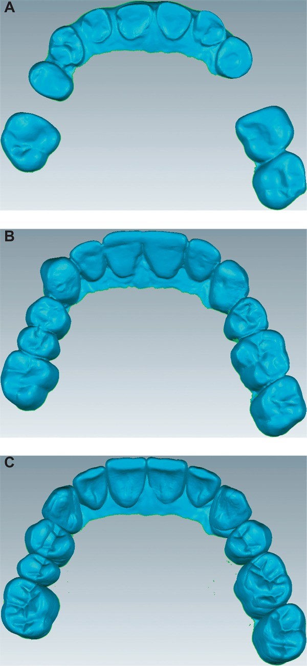
The ideal lateral occlusion scheme has been a subject of considerable discussion in dentistry for many years. It is believed that the lateral occlusion scheme influences masticatory function, comfort, and aesthetics. Altering tooth morphologies, alignments, and orientations during prosthodontic treatment can modify the lateral occlusion scheme. Two common schemes are canine-guided occlusion and group function occlusion. Understanding What Is Canine Guided Occlusion is crucial for effective dental treatment planning.
Understanding Canine-Guided Occlusion
Canine-guided occlusion is a mutually protected occlusion where the vertical and horizontal overlap of the canine teeth causes disengagement of the posterior teeth in the lateral movement of the mandible. It’s speculated that canine-guided occlusion protects the posterior teeth laterally because of the canines’ strategic location, anatomy, and proprioceptive properties.
Objective of Studying Lateral Occlusion
This study aims to evaluate the impact of conventional and digital prosthodontic planning on the lateral occlusion scheme. By understanding how different planning methods affect lateral occlusion, dentists can better tailor treatments to improve patient outcomes.
Materials and Methods Used in the Study
Dental models of 10 patients with Angle Class I occlusion were collected. These patients were undergoing prosthodontic treatment that would influence the lateral occlusion scheme. Each set of models received both conventional and digital wax-ups. Key variables evaluated included:
- Prevalence of different lateral occlusion schemes
- Number of contacting teeth
- Percentage of each contacting tooth
Four excursive positions on the working side were analyzed: 0.5, 1.0, 2.0, and 3.0 mm from the maximal intercuspation position.
Results of the Study
The study found that the lateral occlusion scheme of the two wax-up models was altered following excursion. There was a tendency for the prevalence of canine-guided occlusion to increase, while the prevalence of group function occlusion decreased with increasing excursion. The number of contacting teeth decreased with the increasing magnitude of excursion.
Specific Findings
- At 0.5 mm and 1.0 mm positions, the two wax-ups had significantly greater contacts than the pre-treatment models.
- At 2.0 mm and 3.0 mm positions, all the models were similar.
- Canines were the most commonly contacting teeth, followed by adjacent teeth.
- No difference was observed between the two wax-ups in relation to the number of contacting teeth.
Lateral Movement Simulation
Virtual simulation of lateral movement was performed to understand the occlusal contacts at different excursive positions. This involved moving the mandibular arch horizontally for each specified location and gradually moving the mandible vertically away from the maxilla to detect the existing working side contacts that dictate the lateral occlusion scheme.
Occlusion Schemes Considered
Three occlusion schemes were considered:
- Canine-guided occlusion
- Group function occlusion
- Single tooth-guided occlusion
Prevalence of Lateral Occlusion Scheme
As the excursion increased, the lateral occlusion scheme changed for all evaluated models. The pre-treatment models showed a minimal increase in canine-guided occlusion through the excursion. The group function occlusion tended to reduce with increased lateral excursion.
For the two wax-up models, there was a consistent and gradual increase of canine-guided occlusion and a reduction of group function occlusion with increased excursion. The overall patterns for the two wax-ups were similar, except that the conventional wax-up had a slightly more even pattern of alteration, while the digital wax-up exhibited steeper occlusion alterations.
Percentage of Contacting Teeth
For all arches of all models, the canine had the tendency to be the dominant contacting tooth. Contacts decreased gradually from canines to the more anterior teeth and from the canines to the posterior teeth.
Discussion of the Findings
This study indicates that conventionally and digitally planned prosthodontic treatment influences the lateral occlusion. Multiple lateral locations were considered to provide insight about the possible contact pattern from the immediate excursion to the maximal excursion.
Clinical Relevance
Guiding contacts at the 3 mm position might occur primarily during parafunctional activity and bruxism in the edge-to-edge position. Occlusal guiding during mastication and physiological movement tends to occur within 0.5 mm from the maximal intercuspation. Therefore, the range evaluated in this study covers functional and parafunctional jaw movement.
Dynamic Nature of Lateral Occlusion
The dynamic nature of the lateral occlusion scheme at the different arch positions is attributed to teeth morphological factors. In the initial phase of excursion, the cusps are articulated against wider fossa surfaces. As excursion progresses, the total contact area is reduced, which means fewer teeth will be in contact.
Conclusion
The prosthodontic planning influenced the pattern of the lateral occlusion scheme, with a more prominent influence at the initial stages of excursion. The initial phase of excursion involved a greater number of contacting teeth and a higher prevalence of group function occlusion than maximal excursion. Canine-guided occlusion tends to be more prevalent at the later stage of excursion. Understanding what is canine guided occlusion and its role in dental health is essential for effective treatment planning and improved patient outcomes.
